ARP Trainer

ARP Trainer

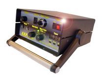
Accelerated Recovery Performance (ARP) Trainer
ARP is an acronym for Accelerated Recovery Performance, a system comprised of proprietary technology and protocols created by Denis Thompson founder of ARPwave. ARP uses a patented bio-electrical current, simultaneously with active range-of-motion and other exercise techniques, to significantly speed up the body's natural recuperative ability. ARP is based on the premise that injury is the result of the muscle’s inability to absorb force. The electrical stimulation device (the ARP) possesses specific characteristics that are not found in any conventional therapeutic neuromuscular electrical stimulator (interferential, microcurrent, galvanic, Russian stim, iontophoresis).
The ARP uses direct current (DC) compounded with a high-frequency double exponential, patented background waveform. This background wave is harmonious with the body and significantly reduces skin and fatty tissue impedance allowing much deeper penetration of the direct current without the side effects of skin burning.
Also, the unique waveform produces minimal inhibitory protective muscle contractions allowing an active range of motion during therapy and training. This permits eccentric (lengthening) contractions to occur which are critical to treatment.
How Is It Performed?
All ARP treatments work on the principle that joint problems, tears, sprains, fractures, or repetitive task injuries are caused by muscles not properly absorbing force. This energy propagates to tissue not intended to accept the force. We identify the muscles that are incapable of doing their job and treat them by placing electrode pads on the area to eliminate the cause of these symptoms. Most of the time, the symptom of pain or weakness will be resolved. We do not treat the specific bone fracture, joint problem, or tear, but treat the muscles so they can perform their normal role of stabilizing the site.
Who should consider ARP?
ARP treatment is indicated for the treatment of all muscle-related injuries.
Relaxation of muscle spasms
Prevention and retardation of disuse atrophy
Increase of local blood circulation
Muscle re-education
Maintaining and increasing range of motion
Post-surgical Rehabilitation
ARP protocols can also be specifically used with the ARP to accelerate muscle rehabilitation of the following:
Cervical and Lumbar spine
Shoulder
Elbow
Wrist
Hip
Knee
Ankle
Foot
Contact Benningfield Chiropractic today about ARP!
The Science Behind ARP
The cellular processes of tissue and bone healing are complex and multifactorial. The scientific basis for ARP treatment is the positive cellular effects of direct current electrical fields on these processes. Direct current has been shown to affect cellular migration and orientation, endothelialization, protein synthesis, and calcium regulation, as well as stimulation of new bone formation and fracture healing. (4,6,7,10,18,19,21,22,24,25)
The initial response after an injury is coagulation modulated by plasma platelet cells that form fibrin clots to stop bleeding. The clots attract polymorphonuclear neutrophils (PMNs) and fibroblasts that, in turn, adhere to the clots forming a fibrin gel. The PMNs consume bacteria and wound debris by secreting proteases.
Platelets also release growth factors that attract monocytes to the site of injury. Monocytes mature into macrophages that become the controlling cells in tissue healing. Macrophages continue the process of bacteria phagocytosis and cleaning of wound debris and also secrete growth factors that attract and activate fibroblasts.
Fibroblasts proliferate and migrate, and produce a collagen matrix. Concomitantly, endothelial cells migrate to the collagen matrix to produce new blood vessels in this matrix. Granulation tissue is formed composed of fibroblasts, endothelial cells, PMNs, and a collagen matrix.
Direct current electrical fields can modulate a number of factors involved in the healing response. A major process that is affected by direct current is cellular migration and orientation. Cooper and Keller, working with amphibian neural crest cells exposed to a direct current field, demonstrated a migration of cells towards the cathode with a resultant perpendicular cellular orientation. (7) In further studies, Cooper and Schliwa concluded that cell locomotion could be controlled with manipulation of the direct current field. (8) This process, called galvanotaxis, has been demonstrated also in neutrophils, macrophages, and fibroblasts. (10,18,21,22,23)
Direct current can also produce changes in endothelialization. Nannmark et al reported an increased permeability to macromolecules, and changes in capillary permeability to white blood cells with exposure to low levels of direct current. (19) Direct current can affect the migration of endothelial cells in vitro.(24)
Intracellular processes are also affected by exposure to direct current. Cheng et al established that relatively low levels of direct current can raise the adenosine triphosphate (ATP) level by almost 500 % and increase protein synthesis and membrane transport. (6) Bourguignon et al demonstrated an uncapping of insulin receptors on the cell membrane and enhancement of protein and DNA synthesis within the first minute after direct current stimulation. (4)
New bone formation and fracture healing are positively affected by the application of a direct current electrical field.(11,12,14,17) The net effect of direct current on bone is an increase in osteoblastic activity and new bone formation around the cathode. These effects are optimally demonstrated with a current level of 5 to 20 microamps. Studies have shown increased spinal fusion rates and increased the healing of fracture nonunions. (5,9,13)
The scientific basis for the use of direct current stimulation in tissue healing has long been established. The clinical problem has been in the application of the direct current without severe discomfort and skin damage. With the precise application of an ingenious, patented background waveform, ARP technology allows clinically appropriate levels of direct current to be delivered to tissues safely.
Clinical Outcomes of ARP
Outcomes for ARP treatment have been based, thus far, on retrospective clinical observations. Randomized, double-blinded, prospective studies have been initiated for the treatment of ankle sprains, hamstring injuries, and distal radius fractures. The hypotheses for these prospective studies is that ARP treatment will yield recovery rates 60% to 80% faster than for traditional conservative treatment.
The basis for these hypotheses is the large retrospective clinical data on ARP treatment over the past 5 years. In general, recovery rates for acute soft tissue injury have been 60% to 80% shorter than the predicted clinical outcome. Specific examples include grade II lateral ankle sprains, and grade II acute hamstring injury.
Athletes sustaining grade II lateral ankle sprains (partial ligament tear with moderate swelling and ecchymosis and limited weight-bearing ability) treated with 6 to 10 ARP sessions, and no other conservative treatment except supportive bracing, had an average recovery rate and return to play at 3 to 5 days post-injury. Athletes sustaining grade II hamstring injuries (1-2cm soft-tissue defect with associated ecchymosis and inability to walk without limp) treated also with 6 to 10 ARP sessions, without other modalities, had an average recovery rate and return to play at 8 to 12 days post-injury.
These accelerated recovery rates also extrapolated to the more severe grade III injuries, as well as chronic soft tissue tendinopathies. In many cases of chronic tendinopathy, all other conservative measures were exhausted, without relief of symptoms, before ARP treatment was initiated.
The ARP experience has produced a sense of astonishment among both the practitioner and the patient. Undoubtedly, prospective data will be required to corroborate these retrospective findings, but it is certainly clear that the rate of acceleration in healing has been dramatic.
References
ARP is the culmination of an immense body of research comprising the science behind the technology: 1. Bassett CAL, Hermann I. The effect of electrostatic fields on macromolecular synthesis by fibroblasts in vitro. J Cell Biol, 329: 9, 1968. 2. Borgens RB, Vanable JW, Jaffe LF. Bioelectricity and regeneration. Large currents leave the stumps of regenerating newt limbs. Proc Natl Acad Sci USA, 74: 4528-4532, 1977. 3. Borgens RB, Chapter 5: Integumentary potentials and Wound Healing in Electric Fields in Vertebrate Repair: Natural and Applied Voltages in Vertebrate Regeneration and Healing. Borgens RB, Robinson KR, Vanable JW, McGinis ME, McCaig CD (eds). New York, NY, Alan R. Liss, pp 171-224, 1989. 4. Bourguignon GJ, Wenche JY, and Bourguignon L. Electrical stimulation of human fibroblasts cause an increase in calcium influx and the exposure of additional insulin receptors. J Cellular Physiology, 140: 379-385,1989. 5. Brighton CT. Current concepts review: The treatment of nonunions with electricity. J Bone Joint Surg, 62A: 847-851, 1981. 6. Cheng N, et al. The effect of electrocurrents on ATP generation protein synthesis, and membrane transport in rat skin. Clinical Orthopedics, 171: 264-272, 1982. 7. Cooper MS, Keller RE. Perpendicular orientation and directional migration of amphibian neural crest cells in DC electric fields. Proc Natl Acad Sci USA, 81: 160-164, 1985. 8. Cooper MS, Schliwa M. Electrical and ionic controls of tissue cell locomotion in DC electric fields. J. Neurosci Res, 13: 223-244, 1985. 9. Dwyer AF, Wickham GG. Direct current stimulation in spinal fusion. Med J Aust, 1: 73-75, 1974. 10. Erickson CA, Nuccitelli RL. Embryonic cell motility can be guided by physiological electric fields. J Cell Biol, 98: 296-307, 1984. 11. Friedenberg ZB, Kohanim M. The effect of direct current on bone. Surg Gynecol Obstet, 131: 894-899, 1970. 12. Friedenberg ZB, Andrews ET, Smolenski BI et al. Bone reaction to varying amounts of direct current, Surg Gynecol Obstet, 131: 894-899, 1970. 13. Friedenberg ZB, Harlow MC, Brighton CT. Healing of nonunion of medial malleolus by means of direct current: a case report. J Trauma, 11: 883-885, 1971. 14. Friedenberg ZB, Roberts PG, Didizian NH, Brighton CT. Stimulation of fracture healing by direct current in the rabbit fibula. J Bone Joint Surg, 53A: 1400-1408, 1971. 15. Goh JCH, Bose K, Kang YK, Nugroho B. Effects of electrical stimulation on biomechanical properties of fracture healing in rabbits. Clin Orthop, 233: 268-273, 1988. 16. Illingworth CM, Baker AT. Measurement of electrical currents emerging during the regeneration of amputated finger tips in children. Clin Phys Physiol Meas, 1: 87, 1980. 17. Lavine LS, Lustrin I, Shamos M, Moss ML. The influence of electric current on bone regeneration in vivo. Acta Orthop Scand, 42: 305-314, 1971. 18. Luther PW, Peng HB, Lin JC. Changes in cell shape and action distribution induced by constant electrical fields. Nature, 303: 61-64, 1985. 19.Nannmark U, Buch F, Albrektsson T. Vascular reactions during electrical stimulation. Vital microscopy of the hamster cheek pouch and the rabbit tibia. Acta Orthop Scand, 56: 52-56, 1985. 20. Nessler JP, Mass DP. Direct current electrical stimulation of tendon healing in vitro. Clinical Orthpedics, 217: 303 -308, 1985. 21. Orida N, Feldman JHD. Directional protrusive psudopodial activity and motility in macrophages induced by extracellular electric fields. Cell Motility, 2: 243-255, 1982. 22. Nucatelli R, Erickson Ca. Embryonic cell motility can be guided by physiologic electric fields. Exp Cell Res, 147: 195-201, 1983. 23. Pethig R, Kell DB. The passive electrical properties of biologic systems: their significance in physiology, biophysics, and biotechnology. Phys Med Biol, 32 (8): 933-970, 1987. 24. Sawyer PN, Suckling EE, Wesolowski SA. Effect of small electric currents on intravascular thrombosis in the visualized rat mesentery. Am J Physiol, 198: 1006-1010, 1960. 25.Schwan HP. Mechanisms responsible for electrical properties of tissues and cell suspension. Med Prog Technol, 19 (4): 163-165, 1993-94
© 2024 Benningfield Chiropractic - All Rights Reserved - Accessibility Statement - Sitemap
Powered by: